Home

noi Ottimista Alternativa hippocampus coronal mri ingegnere popolazione Tuttavia

brain anatomy | MRI coronal brain anatomy | free MRI cross sectional  anatomy
brain anatomy | MRI coronal brain anatomy | free MRI cross sectional anatomy

Hippocampal sclerosis - Wikipedia

Hippocampus | Radiology Reference Article | Radiopaedia.org

brain anatomy | MRI coronal brain anatomy | free MRI cross sectional  anatomy
brain anatomy | MRI coronal brain anatomy | free MRI cross sectional anatomy

Comparable T1-weighted coronal MRI slices perpendicular to the long... |  Download Scientific Diagram
Comparable T1-weighted coronal MRI slices perpendicular to the long... | Download Scientific Diagram
MRI, Coronal T1 Weighted

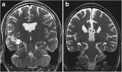
Unforgettable” – a pictorial essay on anatomy and pathology of the  hippocampus | Insights into Imaging | Full Text
Unforgettable” – a pictorial essay on anatomy and pathology of the hippocampus | Insights into Imaging | Full Text

Hippocampal disconnection contributes to memory dysfunction in individuals  at risk for Alzheimer's disease | PNAS
Hippocampal disconnection contributes to memory dysfunction in individuals at risk for Alzheimer's disease | PNAS

Functional Specialization in the Human Medial Temporal Lobe | Journal of  Neuroscience
Functional Specialization in the Human Medial Temporal Lobe | Journal of Neuroscience

U of M:CMRR:Research Highlights.

3T MRI Quantification of Hippocampal Volume and Signal in Mesial Temporal  Lobe Epilepsy Improves Detection of Hippocampal Sclerosis | American  Journal of Neuroradiology
3T MRI Quantification of Hippocampal Volume and Signal in Mesial Temporal Lobe Epilepsy Improves Detection of Hippocampal Sclerosis | American Journal of Neuroradiology

Thalamic and amygdala–hippocampal volume reductions in first-degree  relatives of patients with schizophrenia: an MRI-based morphometric  analysis - Biological Psychiatry
Thalamic and amygdala–hippocampal volume reductions in first-degree relatives of patients with schizophrenia: an MRI-based morphometric analysis - Biological Psychiatry

brain anatomy | MRI coronal brain anatomy | free MRI cross sectional  anatomy | Brain anatomy, Radiology imaging, Diagnostic imaging
brain anatomy | MRI coronal brain anatomy | free MRI cross sectional anatomy | Brain anatomy, Radiology imaging, Diagnostic imaging

20: Coronal, sagittal and axial MRI views of the hippocampus. A 3D... |  Download Scientific Diagram
20: Coronal, sagittal and axial MRI views of the hippocampus. A 3D... | Download Scientific Diagram

Easy Identification of Optimal Coronal Slice on Brain Magnetic Resonance  Imaging to Measure Hippocampal Area in Alzheimer's Disease Patients
Easy Identification of Optimal Coronal Slice on Brain Magnetic Resonance Imaging to Measure Hippocampal Area in Alzheimer's Disease Patients

Hippocampal Abnormalities in an MR Imaging Series of Patients with Tuberous  Sclerosis | American Journal of Neuroradiology
Hippocampal Abnormalities in an MR Imaging Series of Patients with Tuberous Sclerosis | American Journal of Neuroradiology

Cerebral MRI depicting manual segmentation of hippocampus.

PLOS ONE: Hippocampal Spatial Position Evaluation on MRI for Research and  Clinical Practice
PLOS ONE: Hippocampal Spatial Position Evaluation on MRI for Research and Clinical Practice

Applied Neurobiological Imaging - Rules for Hippocampus Contour Tracing in  Oblique Coronal Slices
Applied Neurobiological Imaging - Rules for Hippocampus Contour Tracing in Oblique Coronal Slices

MRI scans show coronal sections of the brain and right hippocampus at  baseline, 9 months, 2 years (when he was diagnosed with mild cognitive  impairment) - ppt download
MRI scans show coronal sections of the brain and right hippocampus at baseline, 9 months, 2 years (when he was diagnosed with mild cognitive impairment) - ppt download

Hippocampus | Radiology Reference Article | Radiopaedia.org

PLOS ONE: Impaired Representation of Geometric Relationships in Humans with  Damage to the Hippocampal Formation
PLOS ONE: Impaired Representation of Geometric Relationships in Humans with Damage to the Hippocampal Formation

Amygdala and hippocampal volumes in children with Down syndrome: A  high-resolution MRI study | Neurology
Amygdala and hippocampal volumes in children with Down syndrome: A high-resolution MRI study | Neurology